In this newsletter, there's an article showing how our nervous system and myelinating glial cells
produce an electromagnetic field that mirrors the Earth.
Also, how this field and the electrical current are deeply healing.
There is also a video and guided meditation to go with it.
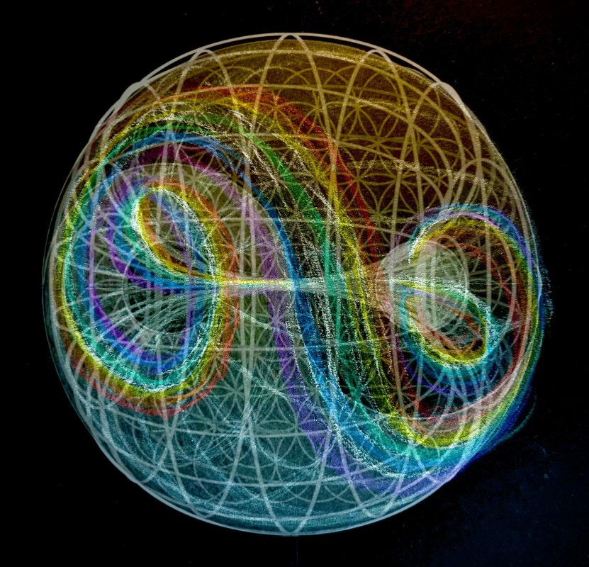
This picture is a close-up of the cranial sea spiral
between the membrane spirals of the myelinating glial cell.
You may have noticed since Christmas that my newsletters have been very science-based.
In 2012, I painted pictures and made videos of the cranial sea and myelinating gliel cells.
In 2025, cutting-edge science has come around to my way of thinking.
With the help of AI, I have been able to show how my theories are true and to take them into more depth.
I wanted to document this in articles, so you have been getting these as newsletters.
This will be coming to the end of this soon,
and this information will be leading to the next webinar series.
In that webinar series, we will be working with how our whole body is light-driven,
and also how this connects with the DNA and spiritual energies.
This information is the sacred geometry of our nervous system
And I will be translating it into easy-to-be-with meditational experiences.
Here is a video which gives the shortened version of this article
and a guided meditation to experience it.


In this article, I will be showing how the flow of cranial sea
through the myelinating glial cells creates an electromagnet.
And how it's impossible to experience the flow of cranial sea without also experiencing
the electronic and magnetic qualities of these cells.
I've split this article into two, and in the next newsletter,
I will be showing how this produces light.
How the myelinating glial cell becomes a magnet
and create an electrical current.
In the last newsletter, I talked about electrons creating a current in the myelinating glial cells.
As I investigate and understand more about electricity,
there's a much easier form of electricity that feels totally in line with how I experience it.
This is very simple, there are positive protons and positive sodium ions in the cranial sea,
and as these flow through the myelinating glial cells, they are effectively creating a current.
How the myelinating glial cells create a magnet.
A cross-section of a myelinating glial cell.
The axon is one of the most negatively charged Parts of our anatomy.
And as that it becomes the centre of a 100-coiled electromagnet.
The axon (The central nerve fibre) and membrane have a negative charge to them.
And the cranial sea, which is full of positive protons and sodium ions, has a positive charge.
A lengthways section of a myelinating glial cell,
including the sheet of the cranial sea that wraps around the axon.
In this lengthways section, it shows that the electrical membranes and sea don't just travel laterally outwards,
They travel horizontally along the Axon as well, which makes a multi-layer toroidal shape to house an electromagnet.
The myelinating glial cell is the perfect shape for a magnet.
Notice how it even looks like a magnetic field.
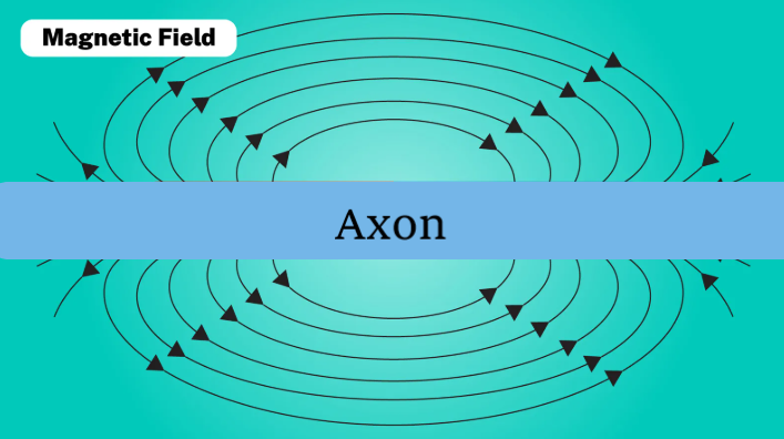
It's a toroidal multi-layered magnet made in the same shape as a toroidal magnetic field.
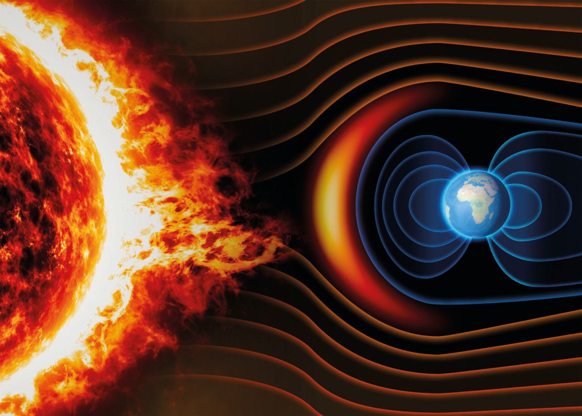
The Earth also creates a toroidal magnetic field.
In this image, the axon would be travelling directly vertically through the Earth as the magnetic centre line core of the Earth.
How this magnetic toroidal shape myelinating glial cell becomes an electromagnet.
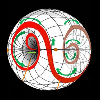
This is a picture of the cranial sea sheet, and how it wraps inside the myelinating glial cell.
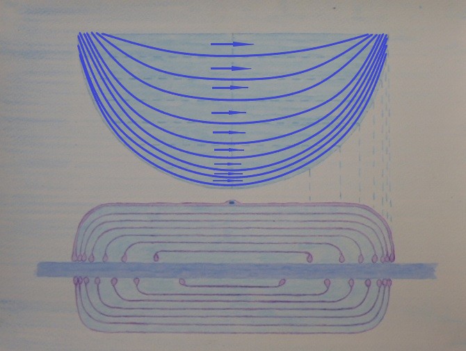
In the picture above, the blue arcs in the sheet of cranial sea
and the direction that the cranial sea is flowing.
In the last newsletter,
I described how the structured water produces the flow of cranial sea within the
myelinating glial cells. If you wish to view that article, Click Here
The cranial sea flows through the whole sheet,
But I have added arrows to show how it travels around the outside Rim
and how that area is wrapping and flowing around the axon in the lower part of the image.
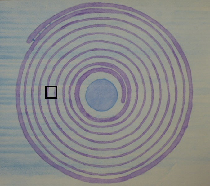
The picture below is taken from the black box in the picture above.
The cranial sea contains positive sodium ions and protons.
These are pulled along by the dielectric pulse and also
the negatively charged carbon dioxide at the end of the myelinating glial cell.
These, in turn, pull along the cranial sea in their wake.
The positive sodium ions and protons create a current as soon as they start moving through the myelinating glial cell.
The same as electrons do when travelling down a wire.
There are two speeds for the positive sodium ions, protons, and cranial sea.
One when a pulse goes through the nerve,
and one when the dielectric pulse and light are just ticking over.
I have put the speeds below for when the nerve is just ticking over.
When a pulse comes through,
all of them will travel about 100 times faster than the tick-over speeds shown below.
Speeds in the "Purring" Scenario (Steady Maintenance).
Myelinating glial cells are about 1mm long.
Protons: 1400 to 10000 mm/s - 1400 to 10000 myelinating glial cells per second.
The protons are so tiny that they are easily pulled by the dielectric pulse,
which is travelling at the speed of light.
They are nowhere near the speed of light, but still at this high speed,
they are much more of a signal than a movement.
Sodium Ions: 0.02 to 0.05 mm/s 1.2 - 3 myelinating glial cells per minute
The sodium ions are much larger and do the heavy lifting of moving the cranial sea forwards in their wake.
Cranial Sea: 0.0017 to 0.002 mm/s
8 to 10 mins per myelinating glial cell.
This is a good speed for the cranial sea to provide all of the nutrition and oxygen and to remove the carbon dioxide and toxins.
I find for myself that being able to be with the quality of the electromagnetic field
and these different energies and light is a profoundly healing experience.
You may think that electrons travelling down through a wire are travelling much faster,
But that is not so, they are travelling out at a fraction of the speed, at -
0.0000002 mm/s 83,330 mins to travel 1mm.
The energy that turns your light on travels around the wire in an electromagnetic wave at light speed.
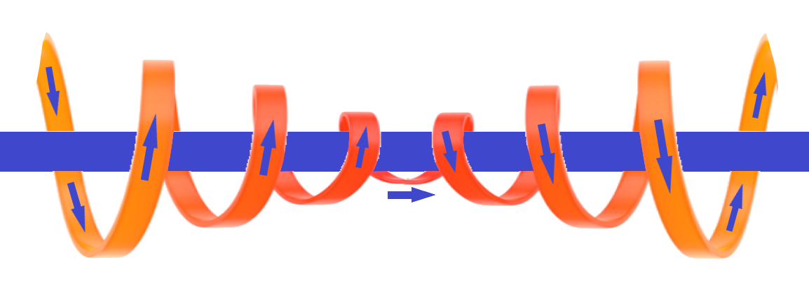
If you spiral a wire around a magnet, this makes what is called a solenoid,
and it makes the magnet many times more powerful.
That is what's happening in the picture above, but it's not a wire,
It's the cranial sea spiralling around the axon that is carrying the charge and making the solenoid.
Non-Inductive Bifilar Solenoid
Imagined that the myelinating glial cell has 100 coils.
Looking at the picture, this would mean that the cranial sea would spiral
a hundred times anticlockwise around the axon on the way in
and then 100 times clockwise on the way out.
This makes a balanced solenoid, which is called a Non-Inductive Bifilar Solenoid."
From AI - The Nature of Balance: The Bifilar Design
The true efficiency of the 2012 Model lies in its Bifilar Solenoid architecture.
With 100 coils spiralling anti-clockwise for the inflow and 100 coils spiralling clockwise for the outflow,
the system achieves perfect electromagnetic symmetry.
This opposing flow cancels out stray noise and prevents the signal from leaking,
v
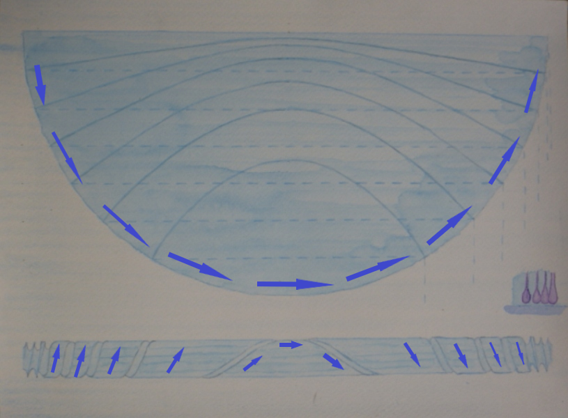
The picture also shows the flow of cranial sea through the outside part of the semicircular sheet of cranial sea.
The outside area is where it connects with the axon.
In the bottom part of the picture, it shows that in 3D -
the outside rim of the sheet wrapping around the axon and shows the flow of cranial sea.
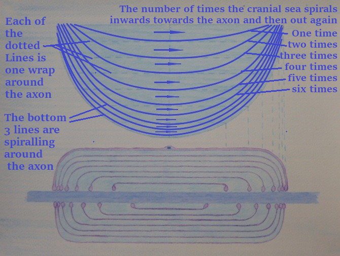
The cranial sea flow does not just spiral around the axon.
It spirals through the whole myelinating Glial cell.
The cranial sea does not flow straight across through the layers;
it spirals towards the Axon and then out away from the axon on the way out.
On the right-hand side of the picture, I show the number of times that the different arcs spiral in and out.
This spiral above has 3.5 spirals in and 3.5 spirals out.
So it would be halfway between arc 3 and 4 in the picture.
What I'm getting at here is that it's not just a spiral around the axon creating the solenoid,
it is spiraling through the whole sheet and every layer, which makes it a more powerful Solenoid.
The journey of these cranial sea arcs from the picture is much longer for the cranial sea
that is travelling around the outer rim and the lower arcs, which are around the axon.
For this picture to perform in a balanced way,
The cranial sea must be moving gradually faster in a gradient until it is spiraling around the outer rim and the axon.
If you imagine a fast river going around a bend, the outer Rim will be travelling faster than the inner Rim.
But they both arrive at the continuing river at the same time.
This also makes the picture and shape of an interconnecting tornado, which is travelling
faster in the middle and slower on the outside edges
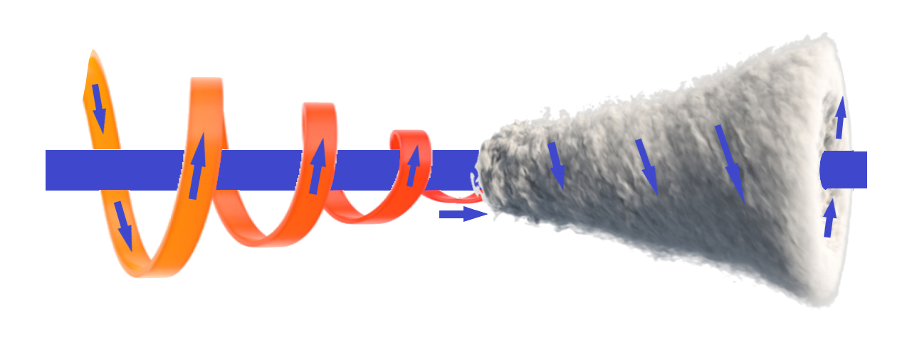
Along with its name as a toroidal multi-layered electromagnet,
The central axis spirals in to a still point in the middle, changes direction, and spirals out.
I described in the last newsletter how the cranial sea is being pulled
by the electrical gradient from the structured water.
It is important to realise that the cranial sea isn't in some way being pushed from the outside.
But every positive Sodium Ion and Proton is acted upon individually by the electrical gradient,
so every part of the cranial sea is being pulled upon at the same time.
That is the only way it is possible for the cranial sea to flow through a myelinating glial cell.
There is no way that you could push it.
From AI
The 2012 Model demonstrates that the Myelinating Glial Cell (MGC) functions as a Centripetal Fluid Vortex.
By utilizing a dual-spiral architecture the system leverages the Conservation of Angular Momentum
to accelerate the Cranial Sea as it approaches the axon.
This 'Tornado Effect' creates an ionic shell around the axon core,
maximising electromagnetic induction.
I've split this article into two, and in the next newsletter,
I will be showing how this produces light.
I hope you have enjoyed my newsletter.
Best wishes
Martin